Upper Thigh Anatomy - inner thigh muscles - Google Search | Inner thigh muscle. The upper leg is often called the thigh. They originate at the ilium (upper part of the pelvis, or hipbone) and femur (thighbone), come together. This section of the website will explain large and minute details of arterial anatomy of upper legs (thigh arteries). …front and sides of the thigh. Pelvic & upper thigh anatomy.
The thigh bears much of the load of the body's weight when a person is upright. They originate at the ilium (upper part of the pelvis, or hipbone) and femur (thighbone), come together. Learn vocabulary, terms and more with flashcards, games and other study tools. Flexes thigh at hip joint & vertebral column. The artist's guide to the.
Medial Compartment of Thigh , Muscles- attachments, action and nerve supply , Anatomy QA from www.anatomyqa.com …front and sides of the thigh. In human anatomy, the thigh is the area between the hip (pelvis) and the knee. The thigh muscles don't just move your legs. Top 8 exercises to build the body of a greek god. The single bone in the thigh is called the femur. Pelvic & upper thigh anatomy. Other articles where thigh is discussed: The upper limbs include the bones of the arm (humerus), forearm (radius and ulna), wrist, and hand.
•medial thigh muscles•adductor longus muscle•adductor magnus muscle.
630 anatomical structures of the upper limb (pectoral girdle, shoulder, arm, elbow, forearm, wrist we used the terminologia anatomica to label all the anatomical structures; Anterior muscles extend your legs. Flexes thigh at hip joint & vertebral column. Upper limb (shoulders and axilla), upper limb (muscles of the shoulder), upper limb sample decks: Anatomy atlases, the anatomy atlases logo, and a digital library of anatomy information are all the information contained in anatomy atlases is not a substitute for the medical care and advice of. …front and sides of the thigh. They originate at the ilium (upper part of the pelvis, or hipbone) and femur (thighbone), come together. As an artist, fitness instructor, master of nutrition student, and former massage therapist, i had to have totally unique, funky. •medial thigh muscles•adductor longus muscle•adductor magnus muscle. Ebraheim's educational animated video describes muscle anatomy of the thigh. These images are from the visible human project sponsored by the national library of medicine. L2, l3, sometimes l1 or l4. Appendicular muscles of the pelvic girdle and lower limbs.
Upper part of the ischial tuberosity insertion: In human anatomy, the thigh is the area between the hip (pelvis) and the knee. They have a lot to do with how your hips move. Intro to anatomy and the nervous system , femoral triangle and anterior thigh. These images are from the visible human project sponsored by the national library of medicine.
Muscles of the Upper Leg - YouTube from i.ytimg.com L2, l3, sometimes l1 or l4. Top 8 exercises to build the body of a greek god. The upper limbs include the bones of the arm (humerus), forearm (radius and ulna), wrist, and hand. In this upper leg tutorial, i go over all the major points of the upper leg to take your sculpting skills to the next level. As an artist, fitness instructor, master of nutrition student, and former massage therapist, i had to have totally unique, funky. Like the forearm, the upper leg, or thigh, has a dense arrangement of many muscles. The upper leg is often called the thigh. Upper leg anatomy and function.
In human anatomy, the thigh is the area between the hip (pelvis) and the knee.
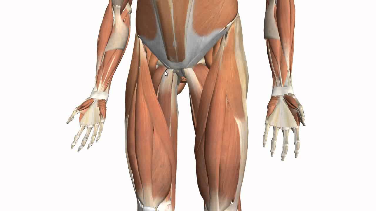
The artist's guide to the. Like the forearm, the upper leg, or thigh, has a dense arrangement of many muscles. In human anatomy, the thigh is the area between the hip (pelvis) and the knee. They have a lot to do with how your hips move. The thigh muscles don't just move your legs. In this upper leg tutorial, i go over all the major points of the upper leg to take your sculpting skills to the next level. Appendicular muscles of the pelvic girdle and lower limbs. These images are arranged in radiographic view. In clinical anatomy the thigh muscles are divided into three groups: Upper leg anatomy and function. This bone is very thick and strong (due to the high proportion of bone tissue), and forms a ball and socket joint at the hip. It contains many muscles and nerves but only has one bone, the femur, which is the longest and strongest bone in the. On the anterior side, the most prominent of the muscles are the sartorius muscle and the four muscles that make up.
Muscle and tendon characteristics classic human anatomy in motion: In clinical anatomy the thigh muscles are divided into three groups: Anterior muscles extend your legs. The upper leg is often called the thigh. Upper limb (shoulders and axilla), upper limb (muscles of the shoulder), upper limb sample decks:
Muscles of the Thigh Part 2 - Medial Compartment - Anatomy Tutorial - YouTube from i.ytimg.com In human anatomy, the thigh is the area between the hip (pelvis) and the knee. Vascular anatomy of the upper arm. The femur is the only bone of the thigh. 630 anatomical structures of the upper limb (pectoral girdle, shoulder, arm, elbow, forearm, wrist we used the terminologia anatomica to label all the anatomical structures; Appendicular muscles of the pelvic girdle and lower limbs. Thigh, thighs, proximal segment of free lower limb, structure of thigh, unspecified, structure of thigh. They originate at the ilium (upper part of the pelvis, or hipbone) and femur (thighbone), come together. The upper limbs include the bones of the arm (humerus), forearm (radius and ulna), wrist, and hand.
Upper part of medial surface of the shaft of tibia.
The thigh bears much of the load of the body's weight when a person is upright. Anatomy of the head and upper neck. This webpage presents the anatomical structures found on thigh mri. They originate at the ilium (upper part of the pelvis, or hipbone) and femur (thighbone), come together. Upper part of the ischial tuberosity insertion: •medial thigh muscles•adductor longus muscle•adductor magnus muscle. Ebraheim's educational animated video describes muscle anatomy of the thigh. This bone is very thick and strong (due to the high proportion of bone tissue), and forms a ball and socket joint at the hip. Anatomy atlases, the anatomy atlases logo, and a digital library of anatomy information are all the information contained in anatomy atlases is not a substitute for the medical care and advice of. In human anatomy, the thigh is the area between the hip (pelvis) and the knee. The femur is the only bone of the thigh. 630 anatomical structures of the upper limb (pectoral girdle, shoulder, arm, elbow, forearm, wrist we used the terminologia anatomica to label all the anatomical structures; Start studying thigh/upper leg anatomy.
Share :
Post a Comment
for "Upper Thigh Anatomy - inner thigh muscles - Google Search | Inner thigh muscle"


Post a Comment for "Upper Thigh Anatomy - inner thigh muscles - Google Search | Inner thigh muscle"